Biliary Tract Cancer: From First Symptoms to Treatment Decisions
Cholangiocarcinoma, gallbladder cancer and ampullary cancer explained for patients: the three-step diagnostic work-up, when jaundice needs drainage before surgery, and current surgery and chemotherapy options, based on the Japanese JSHBPS guidelines (3rd edition).
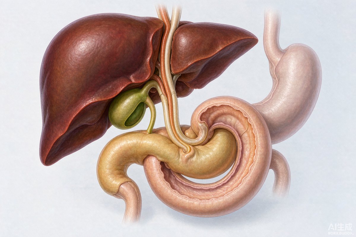
Biliary tract cancers include cholangiocarcinoma (bile-duct cancer, arising anywhere from the tiny ducts inside the liver down to the bowel), gallbladder cancer and ampullary cancer at the drainage opening of the bile duct. They are uncommon but demanding cancers, where the quality of the diagnostic work-up and surgical planning determines outcomes. This guide summarises the Japanese Society of Hepato-Biliary-Pancreatic Surgery (JSHBPS) clinical practice guidelines, third English edition — one of the most widely referenced biliary cancer guidelines worldwide.
Symptoms and risk factors
Most patients notice jaundice — yellowing skin and eyes, dark urine, pale stools — sometimes with pain under the right ribs, nausea or unexplained weight loss. Some cancers are found incidentally on an ultrasound done for other reasons, or in a gallbladder removed for stones. Recognised risk factors include pancreaticobiliary maljunction (a congenital misalignment where pancreatic and bile ducts join too early, allowing digestive enzymes to reflux into the bile duct), primary sclerosing cholangitis and long-standing gallstones.
The three-step diagnostic staircase
Step one is blood testing plus abdominal ultrasound — ultrasound is a strong first test because it is sensitive for bile-duct dilation and gallbladder lesions. Step two is contrast CT and MRI with MRCP (magnetic resonance cholangiopancreatography), which map the tumour's location and spread without invasive tubes. A critical rule in the guideline: CT and MRI must be done before any biliary drainage is placed, because drainage distorts the images. Step three, where needed, is direct examination: ERCP (endoscopic retrograde cholangiopancreatography) allows X-ray imaging of the ducts and tissue sampling; endoscopic ultrasound (EUS) precisely assesses depth of invasion and nearby vessels; and peroral cholangioscopy sends a mini-camera into the duct itself. PET-CT contributes mainly to detecting lymph-node and distant spread. About 69% of patients have an elevated CA19-9, but no blood test is specific.
Prevention: when organs are removed before cancer appears
The guideline strongly recommends preventive surgery for two groups. People with pancreaticobiliary maljunction without bile-duct dilation should have the gallbladder removed (gallbladder-cancer risk as high as 88%); those with dilation should have both gallbladder and the dilated bile duct removed. Sessile gallbladder polyps of 10 mm or more, or polyps growing quickly, are also recommended for surgery. Asymptomatic gallstones alone, however, do not justify removing the gallbladder.
Surgery: the only curative route
If evaluation — including liver-function tests such as the indocyanine green retention test (ICGR15) and CT measurement of the future remnant liver volume — shows the tumour can be removed with adequate margins and safe liver reserve, surgery offers the only chance of cure. Ampullary and lower bile-duct cancers are treated with pancreaticoduodenectomy (Whipple procedure). Hilar cholangiocarcinoma usually requires major hepatectomy; when the planned resection removes half or more of the liver, portal vein embolisation is performed weeks beforehand to grow the remaining side. When jaundice is present before a major liver resection, preoperative biliary drainage — preferably endoscopic drainage placed on the side of the liver that will remain — is recommended. When gallbladder cancer is suspected, open rather than laparoscopic cholecystectomy is advised. The guideline also recommends that hepatectomy and pancreaticoduodenectomy be performed at high-volume centres, where complication and mortality rates are demonstrably lower.
When surgery is not possible
For unresectable obstruction, endoscopic stenting restores bile flow: covered metal stents for lower ducts, plastic or uncovered stents for hilar tumours. First-line chemotherapy is gemcitabine plus cisplatin (or gemcitabine plus S-1); after progression, fluoropyrimidines, and — for the small group with high microsatellite instability — pembrolizumab. Radiotherapy can prolong stent patency and relieve pain. When even these are not tolerated, structured palliative care focused on comfort is the guideline-recommended path.
For international patients
China's major hepatobiliary centres — Zhongshan Hospital and the Oriental Hepatobiliary Surgery Hospital in Shanghai among them — perform some of the highest volumes of biliary surgery worldwide. Two practical points from the guideline matter if you travel for care: bring your CT/MRI on disc rather than only reports, because resectability judgements depend on raw images, and expect the first visit to focus on confirming whether drainage and stenting were placed in a way that keeps surgical options open.