Non-Muscle-Invasive Bladder Cancer: After the First Tumour Is Removed
Why a "superficial" bladder tumour is not the end of treatment, how risk is stratified, what BCG and intravesical chemotherapy do, and how follow-up cystoscopy works — based on the 2021 EAU guideline on Ta, T1 and carcinoma in situ.
Most bladder cancers are diagnosed at a stage where the tumour has not grown into the muscle wall of the bladder. That is genuinely good news, but it is not the end of the story: these tumours recur frequently and a proportion progress to muscle-invasive disease, which requires far more aggressive treatment. The whole strategy of care is therefore built around complete removal, risk stratification, and additional therapy inside the bladder to prevent recurrence and progression. This guide follows the 2021 European Association of Urology guideline for Ta, T1 tumours and carcinoma in situ.
Diagnosis and the first operation
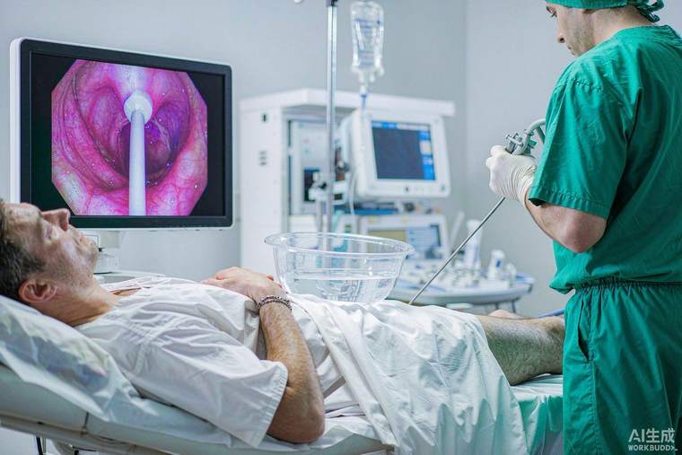
Blood in the urine — visible or found on a urine test — is the usual trigger. Investigation includes urine cytology, imaging of the upper urinary tract, cystoscopy, and often a blue-light or narrow-band imaging technique that improves detection. The first operation is a transurethral resection of the bladder tumour (TURBT), which both removes the visible tumour and provides the tissue the pathologist needs to stage it. Quality matters: the specimen should include muscle in the sample, because without it the pathologist cannot tell whether muscle invasion is present, and a repeat resection is recommended within a few weeks for high-risk or incompletely resected tumours and for T1 disease.
Risk stratification
Patients are grouped as low, intermediate or high risk according to the number and size of tumours, whether they have recurred before, the stage (Ta versus T1), the grade, the presence of carcinoma in situ, and variant histologies. This grouping determines everything that follows. Low-risk disease needs a single immediate instillation of chemotherapy and cystoscopic follow-up. Intermediate-risk disease receives intravesical chemotherapy or BCG for a defined course. High-risk disease — T1, high grade, carcinoma in situ, or multiple and recurrent — receives intravesical BCG, typically for one year of maintenance, because BCG reduces both recurrence and — importantly — progression.
Treatment inside the bladder
A single dose of chemotherapy — mitomycin C, epirubicin or gemcitabine — instilled into the bladder within hours of surgery reduces recurrence after TURBT. BCG is a live attenuated tuberculosis vaccine instilled into the bladder, where it provokes a local immune response against tumour cells; it is given weekly for six weeks as induction, followed by maintenance instillations over one to three years depending on risk. Side effects are mostly local — frequency, burning, blood in the urine — and occasionally fever or a systemic reaction requiring temporary treatment. Alternatives for BCG-unresponsive disease include intravesical chemotherapy combinations, device-assisted treatments, systemic immunotherapy with checkpoint inhibitors, and — frequently the best option — early radical cystectomy.
Carcinoma in situ
Carcinoma in situ is a flat, high-grade lesion that may not form a visible lump; it is detected by cytology, by targeted biopsies or by enhanced imaging. Because it carries a high risk of progression, it is treated with a full course of BCG and followed closely; persistent CIS after adequate BCG is an indication to discuss removal of the bladder.
Follow-up
Surveillance is by cystoscopy at intervals determined by risk — typically at three months initially, then at lengthening intervals for years, since late recurrences occur — often combined with urine cytology and upper-tract imaging in higher-risk patients. Stopping smoking is the single most effective thing a patient can do: it reduces recurrence and progression. Useful questions: was muscle present in my specimen, was a repeat resection advised, what is my risk group, and how long will BCG maintenance continue?