Kawasaki Disease: Protecting the Coronary Arteries
Why every child with Kawasaki disease needs heart scans, how coronary lesions are graded and treated, and how long follow-up lasts — based on China's 2020 revised recommendations.
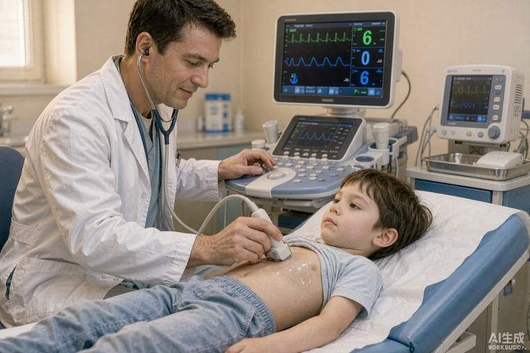
Kawasaki disease is an acute inflammation of blood vessels that almost always affects children under five. Its importance lies in one complication: inflammation of the coronary arteries, which can leave aneurysms that may narrow, clot or, rarely, rupture years later. With prompt treatment the risk of lasting damage drops sharply — which is why timing matters more than anything else. This guide summarises the 2020 revision of China's clinical recommendations for Kawasaki disease coronary artery lesions.
Recognising Kawasaki disease
The diagnosis is clinical: fever lasting five days or more, plus four of five features — changes in the lips and mouth (cracked lips, strawberry tongue), a rash, redness of both eyes, swelling and later peeling of the hands and feet, and swollen neck glands. Incomplete cases exist, especially in infants under six months, who may have fewer signs but a higher risk of coronary injury. Any child with prolonged fever and several of these features should be assessed urgently by a paediatrician.
The treatment window
Standard treatment is intravenous immunoglobulin (IVIG) at 2 g/kg in a single infusion, ideally within the first ten days of fever, together with aspirin. IVIG within this window reduces coronary aneurysm risk from roughly 20–25% to under 5%. About 10–20% of children remain febrile after the first dose; a second dose, corticosteroids or biologic agents such as infliximab are then considered. Aspirin is continued at a low dose afterwards to reduce clotting risk while the arteries heal.
How coronary lesions are graded
Echocardiography is the mainstay and is repeated regularly. Lesions are described as dilation, small, medium or giant aneurysm (a giant aneurysm is generally 8 mm or more in internal diameter). Risk stratification drives treatment: small lesions in the first two months need antiplatelet therapy alone; medium aneurysms usually add a second antiplatelet drug; giant aneurysms require anticoagulation — warfarin or low-molecular-weight heparin — combined with antiplatelet therapy, because the risk of clot inside the aneurysm is high.
Beyond ultrasound
Echocardiography is excellent for the main coronary arteries but is operator-dependent and less reliable in older, larger children. CT angiography maps aneurysm shape and calcification, and cardiac MRI or stress testing assesses whether a narrowed segment is actually restricting blood flow to the heart muscle. Invasive angiography is reserved for planning an intervention such as stenting or bypass.
Long-term follow-up
Follow-up is stratified by the worst lesion ever documented. Children whose scans stayed normal throughout generally need echocardiography at intervals during the first year and can then be discharged with routine care. Those with persisting aneurysms are followed indefinitely: regular echocardiography, periodic stress imaging, and — for giant aneurysms — lifelong anticoagulation and cardiology review. Practical points for families: keep every scan and report, ask about activity restrictions and influenza vaccination, and know the warning symptoms of reduced heart blood flow — chest pain, breathlessness, fainting or unusual tiredness on exertion.