A Kidney Mass Was Found: Evaluation and Diagnosis
How a renal mass is characterised by CT or MRI, when biopsy is worthwhile, and how active surveillance, surgery and ablation are weighed — based on the 2021 AUA renal mass and localized renal cancer guideline, part one.
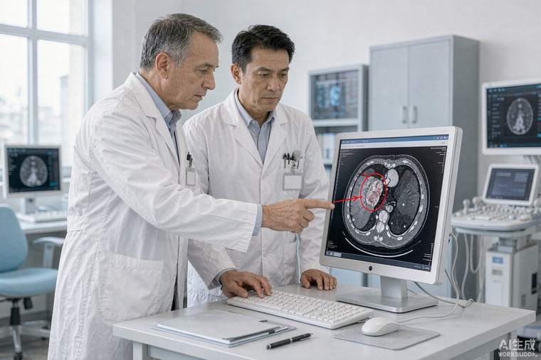
Most kidney masses are now found by chance, on a scan done for something else. That is good news — incidental tumours are usually small and often slow-growing — but it also means the first question is not "how do we remove it" but "what exactly is it, and does it need removing at all?" The 2021 American Urological Association guideline on renal masses and localized renal cancer addresses evaluation, counselling and management.
Characterising the mass
The pivotal test is a dedicated multiphasic CT or MRI of the abdomen with contrast, which shows whether a mass enhances, how it enhances, and how it relates to the collecting system and vessels. Enhancement on contrast imaging is what distinguishes a solid tumour from a cyst. Complex cysts are classified by the Bosniak system (I–IV); Bosniak III and IV cysts carry substantial malignancy risk and are managed like tumours, whereas Bosniak I and II cysts are benign and need no treatment. Ultrasound can identify a mass but cannot reliably characterise it.
Is a biopsy needed?
Renal tumour biopsy has become mainstream and should be discussed rather than assumed unnecessary. It is most valuable when the result would change management: before ablation, before active surveillance, when a metastasis, lymphoma or infection is in the differential, or when the patient's overall condition makes surgery risky. It is not routinely required before surgery in a healthy patient with a typical enhancing tumour. Biopsy is safe, has a low complication rate, and is highly informative when positive — but a non-diagnostic result does not rule out cancer and is generally managed according to the imaging appearance.
Weighing the options
Three strategies exist. Active surveillance — regular imaging to watch the tumour — is an appropriate first choice for many small renal masses, particularly in older patients or those with competing health risks, since most small tumours grow slowly and metastasis while under surveillance is uncommon. Partial nephrectomy removes the tumour while preserving kidney tissue and is preferred whenever technically feasible, because preserving kidney function protects long-term cardiovascular and kidney health. Radical nephrectomy removes the whole kidney and is reserved for large or centrally located tumours, or where partial removal is not safe. Thermal ablation — cryotherapy or radiofrequency — destroys the tumour in place and suits selected small tumours, especially in patients who are poor surgical candidates; it requires a biopsy first.
Counselling points
Decisions should incorporate tumour size, growth rate on prior imaging, location, biopsy result if available, remaining kidney function, age and other illnesses, and patient preference. Ask: is this likely cancer; how fast is it growing; would a biopsy change the plan; what is the chance of preserving the kidney; what does surveillance involve and how often? Genetic evaluation is appropriate for younger patients or those with multiple or bilateral tumours, since several hereditary syndromes cause kidney cancer.