Liver Ultrasound: What the Scan Can — and Cannot — Tell You
A plain-language walk-through of China's 2021 liver ultrasound guideline: how standard scans, contrast-enhanced ultrasound and elastography are used to detect fatty liver, fibrosis, cirrhosis and liver masses, and how often monitoring is needed.
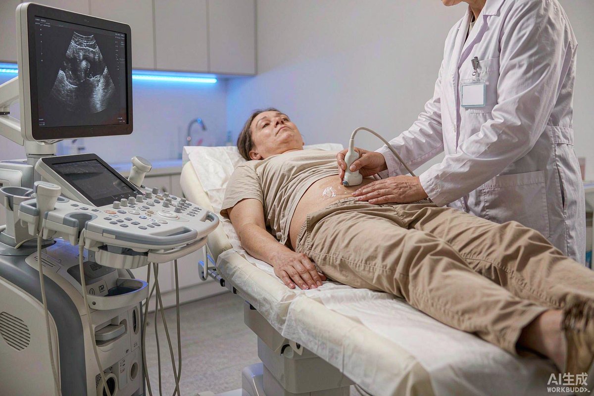
Ultrasound is the world's — and China's — most used liver imaging test: non-invasive, radiation-free, inexpensive and endlessly repeatable. China's 2021 national guideline on liver disease ultrasound standardised how the examination is performed, interpreted and reported. This guide explains what happens during the scan, what the newer ultrasound technologies add, and the monitoring intervals recommended for common liver conditions.
What a standard scan covers
A conventional (grey-scale) scan measures the liver's size and shape, the texture of its tissue, and the calibre and flow of its vessels, alongside the bile ducts and gallbladder. The sonographer works through a defined set of standard views — under the left ribs, below the right ribs and between the right ribs — with you lying on your back or left side. Preparation is minimal: if the bile ducts are part of the question, you fast for 8–12 hours beforehand; a standard liver-only scan usually needs nothing special. The report describes diffuse disease (inflammation, fatty change, fibrosis, cirrhosis) and any focal lesions, mapped to liver segments.
Beyond grey-scale: contrast and elastography
Two newer technologies changed the field. Contrast-enhanced ultrasound (CEUS) injects microbubble contrast through a small IV line and watches how a lesion fills in and washes out in real time. International guidelines now recommend CEUS as the first test when a routine scan finds a liver lump in someone without known cancer and without cirrhosis, when CT or MRI results are inconclusive, and as the preferred contrast method for patients with kidney impairment — the bubbles are safe for the kidneys. Elastography measures liver stiffness, which tracks fibrosis. The two main techniques are transient elastography (FibroScan) and shear-wave elastography on a normal ultrasound machine. As examples of the thresholds used for chronic hepatitis B, stiffness of about 12.4 kPa or more suggests advanced fibrosis, and around 17 kPa suggests cirrhosis; values under 7.4 kPa effectively exclude advanced fibrosis. Fatty change can be quantified at the same time using controlled attenuation (CAP) measurements. The key caveat: stiffness rises with active inflammation and after meals, so elastography is done fasting and rested, and results are interpreted with your blood tests.
Common findings, in plain language
Haemangioma is the most common benign liver tumour — typically a well-defined bright (echogenic) nodule that fills in slowly from the edges on CEUS. Focal nodular hyperplasia is another benign nodule with a central scar and a characteristic spoke-like blood pattern. Simple cysts are trivial. Hepatocellular carcinoma — which develops mostly in cirrhotic livers — shows rapid arterial enhancement with early washout on CEUS. Intrahepatic cholangiocarcinoma tends to wash out even faster, within about a minute. Metastases often appear as multiple nodules with a "target" look. Abscesses evolve over days: early ones mimic tumours, then liquefy, which is why a short-interval repeat scan helps. If you are at risk of liver cancer (cirrhosis or chronic hepatitis B), the report should be read together with your AFP blood test.
How often to scan
The guideline sets explicit monitoring intervals. Chronic liver disease under observation without intervention: abdominal ultrasound every six months. Cirrhosis: ultrasound every six months, plus once-yearly contrast-enhanced ultrasound, CT or MRI to catch hepatocellular carcinoma early. Benign lesions such as haemangioma or focal nodular hyperplasia: yearly ultrasound. After treatment of a liver tumour: every 3–6 months.
For international patients
Liver ultrasound in China is a same-day outpatient study costing a small fraction of Western prices, and top hospitals run dedicated liver units where radiologists, hepatologists and interventionists review images together. Bring previous images and reports — trends over time matter more than any single scan, and the reporting format follows the national guideline described here.