Neurotrophic Keratopathy: When the Cornea Loses Sensation
A degenerative corneal disease caused by damage to the trigeminal nerve — why it is missed, how it is staged, and current treatment options including nerve growth factor — based on a 2021 international expert consensus.
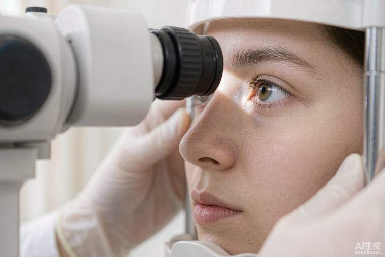
Neurotrophic keratopathy (NK) is a degenerative disease of the cornea caused by loss of its nerve supply — most often the ophthalmic branch of the trigeminal nerve. Without those nerves the corneal surface loses sensation, heals poorly and breaks down, potentially progressing to ulceration, thinning, perforation and permanent vision loss. It is uncommon, frequently under-diagnosed, and dangerous precisely because it causes little pain: patients often present late. A 2021 international expert consensus using a modified Delphi method set out when to screen, how to diagnose and how to treat.
Causes and who should be screened
Recognised causes include herpes simplex and herpes zoster eye infection, diabetes, intracranial tumours or surgery affecting the trigeminal nerve, neurosurgical procedures for trigeminal neuralgia, stroke, and long-term use of preserved eye drops or certain glaucoma medications. Screening is recommended for anyone with these risk factors, and specifically for patients with reduced corneal sensation, unexplained persistent epithelial defects, or a corneal ulcer that does not behave like an ordinary one. The consensus emphasises that NK should be considered in any chronic or non-healing corneal problem, because the treatment is completely different from that of inflammatory or infectious keratitis.
How it is staged
Staging guides treatment. Stage 1 is characterised by surface irregularity, punctate staining and reduced tear stability, with the epithelium largely intact. Stage 2 is a persistent epithelial defect — an area where the surface layer will not heal, often with a characteristic rolled edge. Stage 3 involves stromal involvement: ulceration, thinning, melting, and potentially perforation. Assessment includes measurement of corneal sensation with a Cochet–Bonnet aesthesiometer or a cotton wisp, careful slit-lamp examination, fluorescein staining and photography, and exclusion of infection.
Treatment by stage
Early-stage disease is treated by stopping anything toxic to the surface — preserved drops, unnecessary topical medications — and using unpreserved lubricants, punctal plugs to retain tears, and a therapeutic contact lens or bandage lens to protect the surface while it heals. Infection must be excluded before any steroid is used, and topical steroids are used cautiously because they can worsen melting. For persistent defects and ulcers, options include autologous serum eye drops, amniotic membrane grafting, temporary or permanent tarsorrhaphy (partial closure of the eyelids) to protect the surface, and conjunctival flap surgery in advanced cases.
The specific breakthrough
Recombinant human nerve growth factor (cenegermin) eye drops address the underlying cause rather than the consequences, promoting corneal nerve regeneration and epithelial healing, and have been shown to heal a high proportion of persistent defects and ulcers. The consensus supports its use in moderate to severe disease. Other emerging options include corneal neurotisation — transferring a healthy nerve to the cornea — performed in specialised centres.
Practical advice
Because the eye does not feel pain, the warning signs are visual: blurring, redness, a persistent foreign-body sensation or a white spot on the cornea. Anyone with a history of herpes eye disease, trigeminal surgery or diabetes who notices persistent irritation should have the corneal surface examined. Regular follow-up with photography documents healing, and patients should be told explicitly to avoid preserved drops and to present urgently, not electively, if a defect is found.