NSTE-ACS: Diagnosis and Treatment of Non-ST-Elevation Acute Coronary Syndrome
NSTE-ACS now accounts for over 70 percent of acute coronary syndromes. This guide covers the ECG and troponin strategy, risk stratification, antithrombotic treatment, and when to refer for angiography.
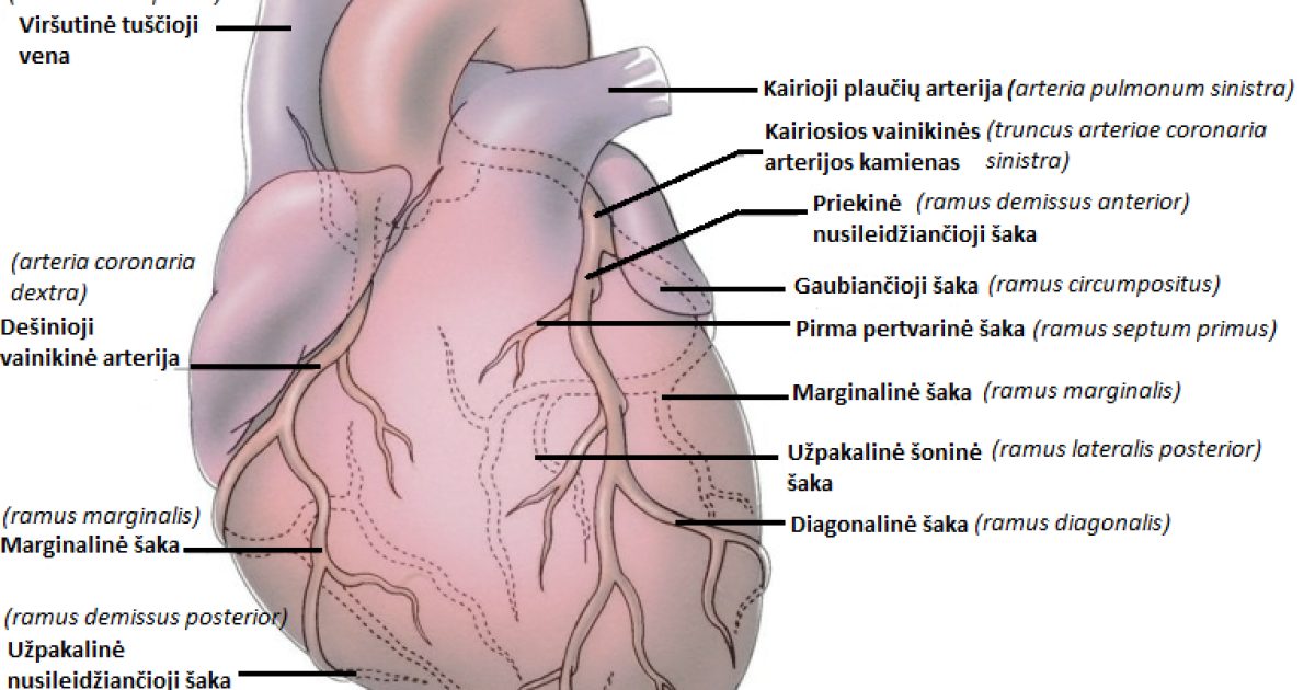
Non-ST-segment elevation acute coronary syndrome - NSTE-ACS - is now the dominant presentation of acute coronary disease, accounting for more than 70 percent of acute coronary syndrome admissions as the incidence of STEMI falls. It comprises non-ST-elevation myocardial infarction, where myocardial necrosis is detectable by troponin, and unstable angina, where it is not. The distinction is a matter of severity along the same pathological spectrum, and the guideline treats them as one condition with a shared diagnostic and treatment pathway.
Although in-hospital mortality is lower than for STEMI, the longer-term picture is worse: rates of reinfarction, readmission and late death are higher, and registry data from China show a substantial burden of events over two years.
Mechanism
Presentation and diagnosis
Rest angina carries a worse prognosis than exertional angina, and angina accompanied by hypotension or heart failure is a particularly adverse sign.
The ECG is recorded within ten minutes of arrival. Diagnostic features are ST depression, transient ST elevation and T-wave changes during pain. If the initial tracing is normal but pain continues, it is repeated within fifteen to thirty minutes, ideally during an episode, and right-sided and posterior leads are added when needed. The number of leads with ST depression and the depth of depression correlate with the amount of ischaemia at risk; ST depression with transient elevation is a high-risk pattern.
High-sensitivity troponin is the preferred biomarker and should be measured within three to six hours of symptom onset. A value above the 99th percentile upper reference limit defines myocardial injury, and the absolute level carries prognostic information. It is important to remember that troponin is not specific to coronary thrombosis - it is also raised in aortic dissection, pulmonary embolism, renal failure, tachyarrhythmias, heart failure, myocarditis and skeletal muscle injury.
Echocardiography assesses left ventricular function and looks for regional wall-motion abnormality, which supports ischaemia. In low or intermediate-risk patients with normal ECG and troponin, provocation testing or coronary CT angiography can exclude the diagnosis.
Differential diagnosis
Treatment
Long-term secondary prevention follows the same principles as for other forms of coronary disease: antiplatelet therapy, high-intensity statin, blood pressure and glucose control, and smoking cessation.