Diagnostic Cerebral Angiography in Children: Why and How It Is Done
When invasive angiography is still needed after MRI and CT, how the procedure is adapted for children, the specific preparation and aftercare, and the risks — based on the 2021 SNIS pediatric practice recommendations.
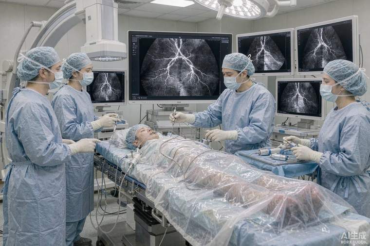
Diagnostic cerebral angiography (DCA) is an invasive X-ray study in which a thin catheter is guided into the neck and head arteries and contrast is injected to show the blood vessels in real time. In children it is no longer a first-line test — MRI and CT angiography answer most questions — but it remains the reference standard when the finest detail of vascular anatomy is needed, and it is the gateway to endovascular treatment in the same session. The 2021 practice recommendations of the Society of NeuroInterventional Surgery (SNIS) set out how the procedure should be adapted for patients under 18.
Why it may be recommended
Typical indications include suspected or confirmed brain arteriovenous malformation, aneurysm, dural arteriovenous fistula, vasculitis affecting the cerebral vessels, certain tumours whose blood supply must be mapped before surgery, and evaluation after head and neck trauma. In many of these, the question is not "is something abnormal" but exactly which vessel feeds it, how fast blood flows through it, and whether adjacent normal vessels are at risk — detail that determines whether, and how, treatment can be given.
Adapting the procedure for a child
Children are not small adults. Considerations include: smaller and more fragile vessels, a higher lifetime stake in radiation exposure, the need for general anaesthesia in most young children (to guarantee stillness and control breathing), different contrast dosing scaled to weight, and a greater sensitivity to blood loss and temperature changes. Radiation is reduced by using low frame rates, tight collimation, fewer projections and paediatric dose settings. Because the physiological reserve is smaller, fluid management, blood pressure targets and temperature control are handled more conservatively than in adults.
Preparation and aftercare
Before the procedure the team reviews kidney function, allergies, blood clotting and current medicines; fasting follows standard anaesthesia rules. Access is usually through the femoral artery in the groin, and ultrasound guidance for puncture is standard practice in children to reduce complications. After the catheter is removed, firm pressure or a closure device seals the artery, and the child lies flat for a monitored period — typically several hours — with regular checks of the puncture site, the pulses in the leg and neurological observations. Most children stay overnight for observation.
Risks, in proportion
Serious complications are uncommon in experienced hands but are real: bleeding or a pseudoaneurysm at the puncture site, injury to the artery or a clot causing stroke, contrast allergy, contrast effects on kidney function, and radiation exposure to skin and, in the lens, to the eyes. Neurological complication rates in published paediatric series are low — generally reported in the low single-digit percentage range including transient events — and are strongly related to operator and centre experience.
Questions worth asking
Is there a non-invasive alternative that would answer the same question? If not, could diagnosis and treatment be combined in one session? How many paediatric angiograms does the team perform each year? What radiation dose-reduction protocol is used? Who will be present — a neurointerventionalist, an anaesthetist with paediatric experience, and nursing staff trained in post-procedure monitoring are the expected minimum. In China, paediatric neurointervention is concentrated in specialised children's and neurosurgical centres, and transferring to such a centre for this procedure is usually worth the journey.