Chest CT in Children with Heart or Lung Disease: When It Is Worth Doing
What cardiothoracic CT adds in congenital heart disease and paediatric chest conditions, how radiation and contrast are handled in children, and how to prepare your child — based on the 2021 Asian Society of Cardiovascular Imaging guidance.
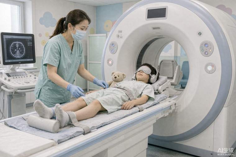
Chest CT produces detailed three-dimensional pictures of the lungs, airways, heart, great vessels and chest wall in a scan lasting seconds. In children with congenital heart disease or complex chest disease, that combination — anatomy, airways and vessels in one dataset — is often what surgical planning requires. The 2021 guidance from the Asian Society of Cardiovascular Imaging congenital heart disease study group focuses on contemporary clinical applications and, importantly, on how to obtain them with the lowest reasonable radiation burden.
When chest CT is chosen
Common indications include defining the anatomy of congenital heart disease before surgery or catheter intervention — especially the pulmonary arteries, pulmonary veins, aorta and collateral vessels; assessing airway compression or tracheobronchial anomalies; evaluating lung parenchymal disease that is unclear on X-ray, such as infection, interstitial disease or congenital lung malformation; and checking for complications such as thrombosis or pulmonary embolism. Echocardiography remains first-line for function and intracardiac detail, and MRI is preferred when a question can be answered without radiation and the child can tolerate a longer scan. CT wins when speed, spatial detail of small vessels, or the airway matters — or when a child cannot lie still long enough for MRI.
Radiation and dose control
Children are more radiosensitive than adults and have more remaining years of life for any risk to express itself, so the guiding principle is "as low as reasonably achievable" while keeping the images diagnostic. Modern practice uses weight- or size-based dose settings, automatic exposure control, low tube voltage appropriate to body size, restriction of the scanned range to the region of interest, and — in cardiac studies — prospective ECG-triggered or high-pitch scanning that avoids continuous exposure. In many paediatric cardiac protocols the dose is now comparable to only a few chest X-rays. Parents should feel able to ask how the dose will be minimised and whether MRI or echocardiography could answer the question instead.
Contrast and preparation
Iodine contrast is given through a cannula, with dose scaled to weight; kidney function is checked beforehand where relevant and hydration is maintained. Timing the scan to the contrast bolus is what makes vessels visible, and in cardiac studies synchronising to the heartbeat is what makes moving structures sharp — sometimes requiring a slower heart rate, though modern scanners tolerate higher rates. Younger children need sedation or general anaesthesia; fasting follows the anaesthetic team's instructions, and a parent may be able to stay during induction.
What the report tells you
Expect a structured description of the heart and vessel anatomy, the airways, the lung tissue, and any incidental findings such as a small nodule or a persistent thymus — the thymus is normal in children and is a frequent source of unnecessary alarm when it is not recognised. Findings are interpreted together with echocardiography, catheter pressures and the clinical picture; a CT does not by itself decide operability. Bring previous imaging and operative notes, because comparison with the child's own earlier studies is often the most informative part of the report.