PET/CT in Childhood Cancer: What Parents Should Know
Why FDG PET/CT is used in children, how the scan is prepared and performed, how radiation exposure is minimised, and what the results can and cannot show — based on the joint SNMMI procedure standard and EANM practice guideline.
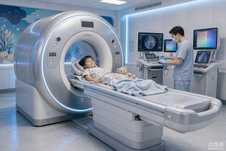
PET/CT with the glucose tracer 18F-FDG shows where cells are consuming glucose unusually actively, which is a hallmark of many cancers. In children it is used to stage disease at diagnosis, to check how well a tumour is responding to chemotherapy, to restage at the end of treatment, and to investigate suspected relapse. The joint SNMMI procedure standard and EANM practice guideline exist to make the study safe, standardised and interpretable — because in children, technique affects both accuracy and radiation dose.
Preparing your child
The tracer is given by injection, so a small cannula is placed first — topical anaesthetic cream helps, and child-life or play specialists are often involved. Fasting for several hours beforehand keeps normal tissues from taking up tracer; water is usually allowed. Blood glucose is checked before injection, since high blood sugar competes with the tracer and degrades image quality. After the injection the child rests quietly for about an hour in a dim room — movement and talking affect uptake, and a calm child produces a cleaner scan. Younger children may need sedation to lie still; the nuclear medicine team will discuss this in advance and give specific fasting instructions if sedation is planned.
The scan itself
The child lies on a padded table that moves through the scanner; the PET acquisition typically takes 20–40 minutes depending on the protocol and the machine, with the CT component used for anatomical localisation and attenuation correction. Modern scanners are designed for paediatrics in two ways: dose-reduction technology, and protocols that scale the injected activity to the child's weight or body surface area rather than using an adult dose. Asking whether the centre uses weight-based paediatric dosing and a low-dose CT protocol is entirely reasonable.
Radiation: putting it in context
Parents understandably worry about radiation. The effective dose from a paediatric FDG PET/CT is real but modest and has fallen substantially with modern equipment — typically a few millisieverts, with the CT component adjusted or, in some protocols, replaced by MRI (PET/MRI) where available. The relevant comparison is clinical benefit: PET/CT frequently changes staging and therefore treatment, and missing active disease carries far greater risk than the scan. Practical safeguards are worth requesting: dose scaled to size, a low-dose CT, shielding where appropriate, and avoiding scans that will not change management.
Reading the results
The report describes tracer-avid sites with standardised uptake values, compares them with prior scans, and flags findings that need confirmation. Important caveats: infection and inflammation also take up FDG, so a positive area is not automatically cancer; some tumours — including several renal and neuroendocrine types and some low-grade brain tumours — take up little FDG and may need MRI or a different tracer; and the thymus or brown fat in children can light up normally. Results should always be interpreted by the treating team alongside MRI, pathology and blood markers. Bring previous imaging on disc so comparisons are like-for-like.