After Kidney Cancer Treatment: Surveillance and What to Watch For
How active surveillance is conducted, the follow-up schedule after surgery or ablation, and how recurrence is detected — based on the 2021 AUA guideline, part two.
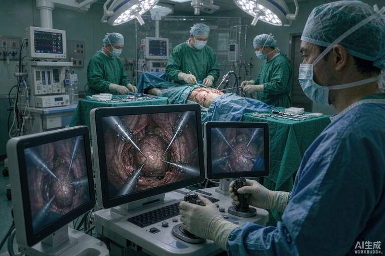
Treatment for a localised kidney tumour is not the end of the story: small tumours can behave unexpectedly, and treated patients remain at risk of recurrence or a new tumour in the same or the other kidney. The second part of the 2021 AUA guideline covers active surveillance and follow-up after intervention, and its practical message is that surveillance is a structured protocol, not an occasional scan.
Active surveillance in practice
For a small renal mass under surveillance, the protocol is regular imaging — usually abdominal CT or MRI at defined intervals, often with an early scan at three to six months to establish the growth rate, then at lengthening intervals if stable — combined with periodic chest imaging and assessment of kidney function. The parameters that matter are size, growth rate (typically expressed as millimetres per year) and any change in appearance. Intervention is triggered by documented growth beyond a threshold, increasing complexity, or patient preference. Biopsy is often incorporated into surveillance, particularly before ablation. Surveillance is safest in patients whose general health would make surgery risky and in those with small tumours; it requires reliability in attending scans, and it carries a small but real risk of progression that should be discussed explicitly before choosing it.
Follow-up after surgery
After partial nephrectomy, follow-up is more intensive than after radical nephrectomy because the remaining kidney tissue could harbour a recurrence at the surgical site; imaging of the operated kidney is therefore part of the schedule. Surveillance typically includes abdominal imaging at intervals over the first few years and periodically thereafter, plus chest imaging, since the lungs are the commonest site of metastasis. Blood tests focus on kidney function — creatinine and estimated filtration rate — and, where relevant, on the metabolic consequences of reduced kidney reserve: blood pressure, proteinuria and anaemia.
Follow-up after ablation
Ablation requires particularly careful imaging review, because a successfully treated tumour leaves a scar that must be distinguished from residual disease. Protocols call for contrast imaging at defined intervals after the procedure, with specific attention to any nodular or peripheral enhancement, which suggests residual tumour rather than expected involution. Biopsy or repeat ablation may follow if findings are equivocal.
Detecting recurrence
Recurrence is most often found on scheduled imaging rather than through symptoms, which is why adhering to the schedule matters more than the individual test. Symptoms that should prompt earlier review include new flank or bone pain, weight loss, persistent cough, blood in the urine, or a lump. Metastases, when they occur, are treated with systemic therapy — targeted agents and immunotherapy have largely replaced older cytokine treatments — and, in selected cases, metastasectomy or stereotactic radiotherapy to isolated deposits.
Living well afterwards
Protecting remaining kidney function is the long-term priority: control blood pressure, avoid routine NSAID use, stay well hydrated, and have kidney function checked at least annually. If you had a partial nephrectomy, ask for your baseline kidney function and a plan for monitoring it. Keep a personal record of scan dates and tumour measurements — a simple table makes growth trends obvious and makes consultations more productive.