Triple-Rule-Out CT: One Scan for Three Chest Emergencies
How a single CT excludes coronary syndrome, pulmonary embolism and aortic dissection in emergency chest pain, what preparation is needed, and the technique factors that affect accuracy — based on the Chinese expert consensus on triple-rule-out CT angiography.
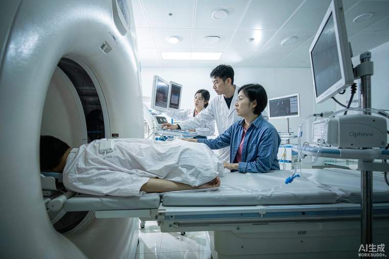
Acute chest pain has three immediately life-threatening causes that need to be distinguished within minutes: acute coronary syndrome (a blocked coronary artery), pulmonary embolism (a clot in the lung artery) and aortic dissection (a tear in the aorta). Triple-rule-out CT angiography is a single contrast-enhanced scan designed to visualise the coronary arteries, pulmonary arteries and aorta at once, avoiding a sequence of separate tests in an emergency department. The Chinese expert consensus on the technique standardises patient preparation, scanning protocol, image reconstruction, quality control and radiation dose.
Who it is for
The scan suits patients with acute chest pain in whom the clinical picture and initial tests — ECG, troponin, chest X-ray, D-dimer — do not clearly point to one of the three diagnoses, and in whom a rapid answer would change management. It is less appropriate when a single diagnosis is already obvious (for example, a classic ST-elevation heart attack goes to the catheter laboratory), when kidney function or contrast allergy precludes contrast, or when the patient is too unstable or arrhythmic for a timed acquisition.
Preparation and scanning
A large-bore cannula is placed, usually in the right arm, and contrast is injected at a high flow rate with the scan timed to the arrival of contrast — synchronisation is what makes the images diagnostic. Heart rate matters for coronary image quality: the consensus advises tailoring the scan mode to the patient's heart rate and the scanner type, generally aiming for a rate below about 70 beats per minute on 64-slice scanners, while dual-source and wide-detector scanners tolerate rates up to about 90. Importantly, beta-blockers are not routinely given beforehand in this emergency setting. Breath-holding for roughly ten to fifteen seconds is required, which is why the team rehearses the breath-hold first.
Quality and dose
Image reconstruction matters as much as acquisition: thin slices are reconstructed and then reviewed as multiplanar reformats and three-dimensional views, because coronary and pulmonary vessels are assessed along their course rather than in a single plane. Radiation dose is controlled by prospectively triggered or high-pitch acquisition where possible, limiting scan range, and using automated exposure control. Quality is judged objectively — adequate contrast opacification and absence of motion artefact in each of the three vascular territories — because a technically inadequate scan is worse than none: it neither excludes nor confirms disease.
What the result means
A negative triple-rule-out CT substantially reduces the probability of the three emergencies, allowing a safe, earlier discharge — but it does not exclude every cause of chest pain, and myocarditis, pericarditis, reflux, musculoskeletal pain and anxiety are outside its scope. Incidental findings are common — small lung nodules, thyroid or liver lesions — and these usually need outpatient follow-up rather than urgent action. If the scan shows one of the three, the emergency team moves directly to the relevant pathway: cardiology, anticoagulation or vascular surgery.
Practical points for patients and families
Tell the team about kidney disease, previous contrast reactions, thyroid disease, metformin use and pregnancy before the scan. Afterwards, drink plenty of fluids unless you have been told to restrict them, and ask specifically whether any incidental finding needs follow-up and when.