Urethral Injury: Diagnosis, Timing and Reconstruction
How urethral injuries are classified and investigated, why immediate repair is not always best, and what reconstruction involves — based on the 2020 European Association of Urology (EAU) guidance on urethral trauma.
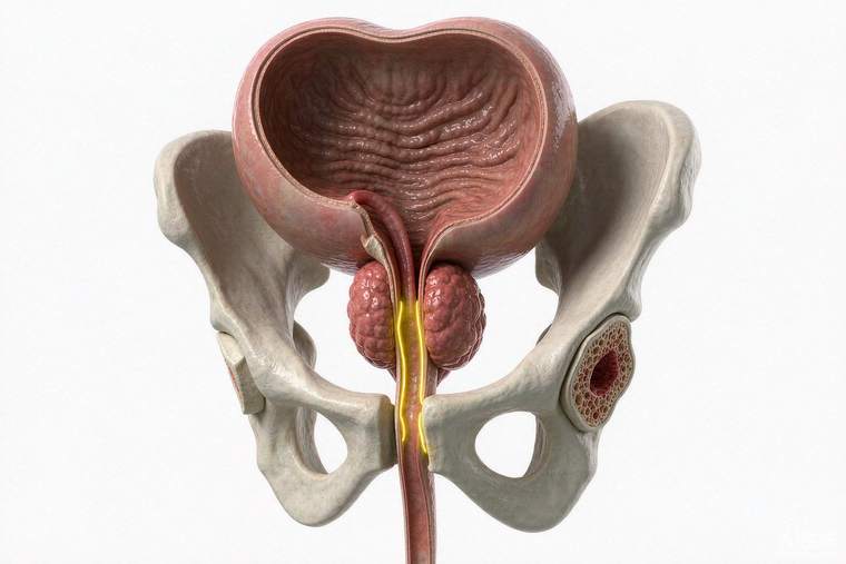
The urethra can be injured by blunt trauma (a straddle injury or pelvic fracture), by penetrating injury, or — most commonly of all — by medical instruments, particularly catheterisation. Iatrogenic injury during catheterisation is reported in roughly six to seven per thousand catheterisations and is usually caused by a false passage, inflating the balloon inside the urethra, or removing a catheter before the balloon is fully deflated. Correct technique prevents most of these. The EAU's guidance on urethral trauma separates anterior from posterior injuries, because their mechanisms, investigation and timing of repair differ.
Recognising the injury
Typical signs include blood at the urethral opening, inability to pass urine, pelvic or perineal pain and bruising, a palpable bladder, and — in pelvic fracture — a prostate that feels high or absent on rectal examination. A key rule: if urethral injury is suspected, do not attempt catheterisation blindly; a suprapubic catheter or specialist-guided placement is safer, and a blind attempt can convert a partial tear into a complete one. Any difficulty passing a catheter, or blood after a difficult catheterisation, warrants urological assessment.
Classification and investigation
Anterior urethral injuries involve the bulbar and penile urethra, typically from a straddle injury crushing the urethra against the pubic bone; about 15% of penile fractures involve the urethra. Posterior injuries involve the membranous and prostatic urethra and are associated with pelvic fractures. Investigation is by retrograde urethrography — contrast gently injected into the urethra under X-ray — which shows whether the urethra is intact, stretched or disrupted, and where; this is often combined with cystography and CT when bladder injury or pelvic fracture is present. Ultrasound and MRI have roles in selected cases, and endoscopy can clarify partial injuries.
Timing: not everything is repaired immediately
For posterior injuries associated with pelvic fracture, the standard approach is early urinary diversion with a suprapubic catheter and delayed reconstruction — typically after three months, once the pelvic haematoma has resolved and the scar is mature — because immediate open surgery in a fresh pelvic haematoma risks bleeding, impotence and incontinence, and the urethra often heals with a treatable short stricture that can be managed later. For anterior blunt injuries, the guidance distinguishes partial from complete disruption: partial injuries can be managed with urinary diversion, whereas complete disruption can be treated with immediate urethroplasty in appropriate hands. Penetrating injuries generally warrant early exploration and repair, along with assessment of the penis, testes and pelvis.
Reconstruction
When a stricture or gap results, urethroplasty is highly successful in experienced hands. Short bulbar strictures are often treated by excision and primary anastomosis; longer or complex strictures use graft (buccal mucosa) or flap techniques, sometimes staged. Success rates are high but recurrence can appear years later, so follow-up includes flow-rate testing, symptom review and, where indicated, imaging or endoscopy.
What to expect afterwards
Recovery involves a catheter for a period, activity restrictions, and follow-up to confirm flow. Erectile and urinary continence outcomes depend on the injury pattern and its treatment — questions to ask before reconstruction are how many of these procedures the surgeon performs, what the expected success rate is for this specific stricture, and how complications such as recurrence or sexual dysfunction are handled.