Bell's Palsy: China's 2022 Guideline on Facial Nerve Repair and Recovery
Most people recover from Bell's palsy, but a third of moderate and severe cases leave lasting problems. The 2022 guideline covers staging, electrodiagnostic testing that predicts outcome, and combined treatment.
Bell's palsy - idiopathic facial nerve paralysis - is the commonest cause of facial nerve weakness, accounting for 60 to 75 percent of cases, with an incidence of roughly 11 to 53 per 100,000. It produces sudden weakness of the muscles on one side of the face and, because it affects expression, it has an outsized impact on how patients see themselves and how they are seen.
The Chinese 2022 clinical guideline on neurorestorative treatment of idiopathic facial palsy sets out how the condition is staged, how prognosis is assessed, and what combination of treatments is recommended.
Why the nerve fails
Presentation
Examination shows a flattened nasolabial fold, a widened palpebral fissure and loss of forehead wrinkling on the affected side. On attempted eye closure the eye rolls upward - Bell's phenomenon.
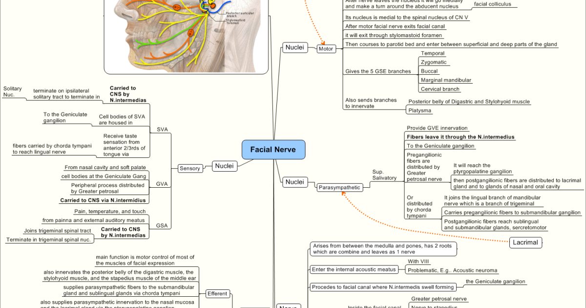
The site of the lesion changes the picture. Proximal to the geniculate ganglion there is loss of taste over the anterior two-thirds of the tongue and hyperacusis. At the geniculate ganglion, herpes zoster infection produces Ramsay Hunt syndrome with vesicles in the external canal and reduced sensation. Near the stylomastoid foramen there is facial weakness plus retroauricular tenderness.
Staging and assessment
Grading uses House-Brackmann, the Burres-Fisch score, and the Sunnybrook system; the Facial Disability Index captures the quality-of-life impact.
Electrodiagnostic testing
Electroneurography compares the compound muscle action potential amplitude with the healthy side and is the best quantitative measure of axonal loss. Degeneration below 90 percent at three weeks supports continued conservative treatment; reaching 90 percent by day six, or a rapid increase of 15 to 20 percent over two days, is an indication to consider surgical exploration.
Needle electromyography is best performed at two to three weeks, when wallerian degeneration is complete; earlier testing under-detects axonal loss. Fibrillation potentials and reduced recruitment denote severe injury, while a recordable evoked potential with normal latency indicates neurapraxia and a good prognosis.