In short
Most pancreatic cysts are harmless and most pancreatic surgery is avoidable. China's 2022 guideline defines which cyst features warrant endoscopic ultrasound, which warrant surgery, and which simply warrant watching.
What this guide covers: The main types · Symptoms are unreliable · Imaging first · Tissue and fluid sampling · Surveillance rather than surgery
Pancreatic cystic neoplasms are being found far more often than they used to be, simply because more people have abdominal imaging. Most of these cysts will never cause a problem, but a minority are premalignant, and telling the two apart is the entire clinical challenge.
China's 2022 diagnostic guideline for pancreatic cystic neoplasms — produced by the National Clinical Research Center for Digestive Diseases with the Chinese Medical Doctor Association pancreatic disease committee, using a modified Delphi process — offers 17 graded recommendations. Its overall message is restraint: distinguish carefully, operate selectively, and avoid over-treatment.
The main types
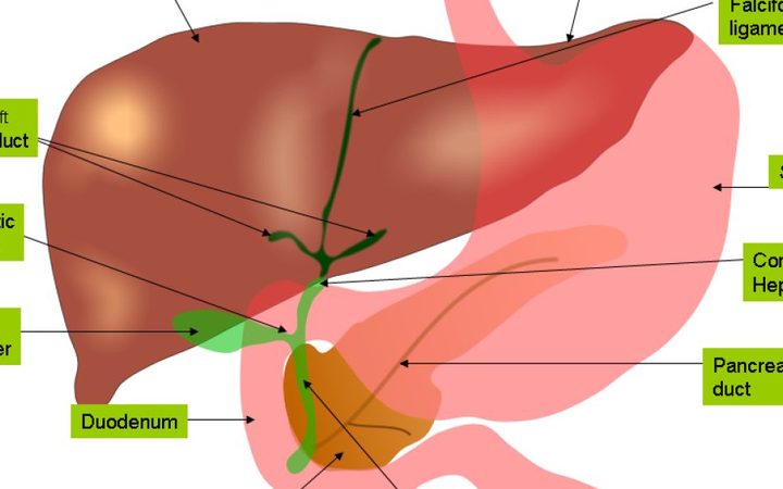
The category includes mucinous cystic neoplasm, intraductal papillary mucinous neoplasm (IPMN), serous cystic neoplasm, solid pseudopapillary neoplasm and cystic neuroendocrine tumour. The distinction matters because mucinous lesions — MCN and IPMN — carry malignant potential, whereas serous cystadenoma is almost always benign.
Symptoms are unreliable
Most patients have no symptoms at all; the cyst is incidental. When symptoms occur they are usually abdominal pain, jaundice or new diabetes. Pain in IPMN often reflects mucus obstructing the main pancreatic duct and causing pancreatitis. Jaundice suggests compression or invasion of the bile duct and generally indicates more advanced disease. The guideline advises caution in attributing vague symptoms to a small incidental cyst, because the association between symptoms and malignancy in IPMN is weak.
Imaging first
MRI is the preferred first-line investigation. It characterises the cyst, shows whether it communicates with the pancreatic duct, and identifies mural nodules and septa — the features that drive risk.
Endoscopic ultrasound is not required for every cyst. It is recommended when imaging shows worrisome features: cyst larger than 3 cm, a mural nodule over 5 mm, thickened or enhancing wall, main pancreatic duct dilatation beyond 5 mm, abrupt duct cut-off with upstream atrophy, lymphadenopathy, rising CA19-9, or growth of 5 mm or more over two years. Combining EUS with cross-sectional imaging improves diagnostic accuracy.
Tissue and fluid sampling
EUS-guided fine-needle aspiration or biopsy is recommended when imaging cannot characterise the lesion or when the result would change management. Cyst fluid analysis — including a string test, cytology, amylase, CEA and glucose, and KRAS or GNAS mutation analysis — helps classify the cyst type. The guideline notes that EUS-guided through-the-needle biopsy outperforms standard FNA for diagnostic yield with a low adverse event rate.
Surveillance rather than surgery
For cysts without worrisome features the recommendation is interval imaging. The purpose of follow-up is twofold: relieve symptoms if they develop, and prevent malignant transformation, while avoiding surgery that the patient does not need.
What patients should ask
Four questions cover it. What type of cyst does the imaging suggest? Are there any worrisome features? Do I need endoscopic ultrasound, or is MRI follow-up enough? And what is the interval for the next scan? A centre following the guideline will have a specific answer to each, and will not recommend resection on the basis of size alone.
Sources and further reading
One reference to a U.S. government health source (National Institutes of Health or MedlinePlus) for the medical background of this guide. It is context, not a substitute for advice from your treating doctor.
General information for international patients, not medical advice or a diagnosis. Read the medical disclaimer.